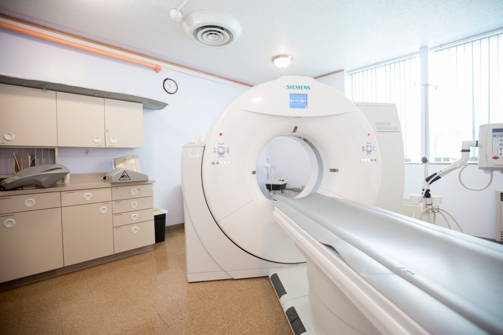
Our radiology department provides comprehensive diagnostic imaging using state-of-the-art equipment and advanced imaging techniques.
Staffed 24 hours a day, 7 days a week, our radiology department offers the following diagnostic tests to our patients.
Computed Tomography (CT Scan)
Mahnomen Health is proud to offer a 20 slice GE scanner. This allows for shorter scan times and shorter breath holds for patients.
What is a CT Scanner?
A CT scan is a specific kind of X-ray. The scanner makes cross sectional images of body tissues.
How is the CT test done?
You may wear your own clothes for the scan as long as there are no metal objects (such as zippers or snaps) near the body part to be scanned. Sometimes a contrast media is used to give more information about your body. If contrast is needed, a nurse will start an IV in a vein, usually in the arm or hand. This involves a small poke using a small IV needle. Once the IV is in place, the needle is removed and a tiny plastic tube stays in the vein during the scan.
The room lights may be dimmed during the scan. If contrast is used, it will be given by IV during the scan. You may experience a warm sensation, a metallic taste in your mouth, or a feeling of needing to urinate as the contrast is given. This is perfectly normal.
You must hold very still and not talk during the scan. You might be asked to hold your breath. We ask that you do the best you can and breathe when the machine tells you to.
What should I do before the CT scan?
You should have nothing to eat or drink at least 4 hours before the scan. If oral contrast is needed, further instruction will be provided by the nurse or radiology technologist.
What should I expect after the CT test?
If IV contrast was administered, it will pass through the kidneys unnoticed in your urine. It’s important to drink extra water after the scan to help pass the contrast. The ordering doctor will contact you with results.
MRI (Magnetic Resonance Imaging)
An MRI scanner creates three dimensional images of body tissues using a large magnet and radio waves. It does not use radiation. There are no known harmful effects from having an MRI scan.
How is the MRI done?
You will go out to the mobile MRI unit. Due to the strength of the magnetic field, all metal objects such as barrettes, bobby pins, jewelry, and credit cards must be removed to give more information about your body. If contrast is needed, an IV will be started in the arm or hand by the MRI technologist.
You will be lying on the imaging bed, which moves in and out of the opening smoothly. A coil will be placed over the part of the body being scanned. The scanner will not touch you, but it will make a loud thumping or tapping sound. You will be given earplugs to wear. You must remain very still during the test.
What should I expect after the MRI?
If IV contrast was injected, drink plenty of water after the test to help flush it through your kidneys. The ordering doctor will contact you with results within a few days.
Echocardiogram
An echocardiogram is a safe and painless procedure that involves sound waves to produce pictures of the heart. The technologist will apply gel to the chest and move a transducer over the area to send sound waves and create images of the heart on the computer. Images are interpreted by Sanford Health.
Digital Mammography
The mammography machine provides exceptionally sharp, digital images with excellent contrast and consistency.
Bone Density (DXA) test
This scan uses small amounts of radiation to determine the density of your bones. This test is used to diagnose osteoporosis or other bone softening. The scan is performed by trained technologists and takes approximately 10 minutes to complete.
Computed Radiography (X-Ray)
An x-ray is a common imaging test that has been used for decades to help doctors view the inside the body. Your doctor may order an x-ray if he or she needs to look inside your body. For example, your doctor may want to view an area where you are experiencing pain, monitor the progression of a disease, or see the effect of a treatment method.
Some conditions that may call for an X-ray include:
- Arthritis
- Blocked blood vessels
- Bone cancer
- Breast tumors
- Conditions affecting the lungs
- Digestive problems
- Enlarged heart
- Fractures
- Infections
- Osteoporosis
- Swallowed items
- Tooth decay
Ultrasound
An ultrasound scan is a medical test that uses high frequency sound waves to capture live images from the inside of your body. An ultrasound allows your doctor to see problems with organs, vessels, and tissue without needing to make an incision. Unlike other imaging techniques, ultrasound uses no radiation, so it is the preferred method for viewing a developing fetus during pregnancy.
How is an Ultrasound done?
The ultrasound technologist will bring you into an exam room, and you will lie on a table. The lights in the room may be dimmed. The technologist will apply warm gel to the transducer and on the area to be scanned. The transducer sends sound waves through the body.
As the sound waves travel through the body, they send echoes back to the computer. The computer makes images of the sound wave patterns. Ultrasounds are usually painless; you may feel slight pressure as the transducer is moved over the skin.
What should I do before an Ultrasound?
Depending on the test ordered, you may be asked not to eat or drink anything including water before the test. A nurse will provide you with those instructions prior to the exam date.
What should I expect after the Ultrasound?
You may go back to normal eating and activity after an ultrasound. You will receive the results from the ordering physician.

Do you have a referral?
Patients at Mahnomen Health are referred to radiology services by a referring physician.
For those patients needing to schedule a mammogram, call (218) 935-2511.